Clemson University professors Richard Figliola and Ethan Kung work on new engineering technologies to perform “virtual vascular surgeries” on patients to help predict surgical outcomes and make surgical decisions before a patient actually goes under the knife. Work they are conducting can mimic a patient’s anatomical geometries as well as overall physiology in order to simulate different cases of surgery at no risk to the patient. “It is possible that in the foreseeable future,” Kung said, “every surgeon will use one, or several, virtual surgeries to practice on a patient before heading to the operating room.”
Like a game of “What If?”, we now have the technology to try out vascular surgeries on patients without actually ever doing anything to them. Two research groups at Clemson University collaborate with a trans-Atlantic network of researchers to tackle complex open-chest surgeries in congenital heart disease this way.
In Mechanical Engineering, Dr. Richard Figliola‘s research group can replicate cardiovascular disease states using physical flow systems. Dr. Ethan Kung‘s research group further utilizes 3D computational fluid dynamics combined with physiology modeling to describe a patient computationally. Using these research tools, surgical treatments and patient responses can be simulated physically and computationally, providing valuable information to a clinician before a treatment decision is made.
Imagine an infant being born with only half a heart– this is essentially the case in single-ventricle heart defect. Without immediate surgical treatment, it is a lethal medical condition. The restoration of the child’s circulation to a final stable state requires several open-chest vascular surgeries spanning over the first few years of the child’s life. At each stage of the surgical treatment, major blood vessels are cut and connected to re-route the circuit for maintaining a delicate balance of blood flow to the lungs and to the rest of the body.
There are many questions associated with these surgical procedures. Where should the blood vessels be cut? Should an artificial graft be used for connecting the vessels, or should we “borrow” tissue from part of the heart itself? What kind of resulting anatomical geometry will be optimal for the patient? These are not questions with clear answers. Traditionally, clinicians have looked to empirical data– experiences accumulated over the years in patient populations, to extrapolate information for any specific patient being considered.
Wouldn’t it be great to find answers to these questions that are specifically tailored to each patient’s unique scenario? The Modeling of Congenital Hearts Alliance (MOCHA) is a group of researchers who have been working together for the past several years to do just that. Funded by the Leducq Foundation, this research network is led by Clemson University and the Great Ormond Street Hospital for Children at the University College of London, and includes engineers and clinicians spanning from Western US, to Italy, France, and England. In this research collaboration, a workflow is created where clinicians provide pre-surgical patient data to engineers, who use it to setup and perform computational and physical modeling of “virtual” surgery of the specific patient, and return the results for clinical consideration.
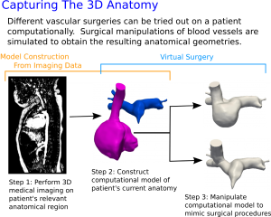
Modern medical imaging modalities such as magnetic resonance imaging and computed tomography provide detailed 3D information of the patient anatomy. Using this imaging data, the researchers computationally construct a 3D representation of the surgically relevant regions. Outside of the 3D region, the researchers use a complex circuit model to represent the patient’s overall physiology. Each circuit model is specifically tuned to describe the characteristics of the unique patient. Clinical information such as pressure, flow, and heart volumes acquired in the patient before a surgical procedure provides insight regarding the patient’s natural physiology and is used to fine-tune the circuit model.
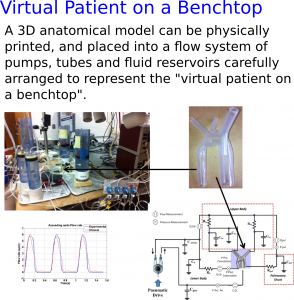
A 3D anatomical model can also be reproduced physically—using what’s called “stereolithography”, or “3D-printing” to obtain a physical model of the anatomy which you can hold in your hands. The researcher can place this physical model into a flow system of pumps, tubes and fluid reservoirs carefully arranged together to represent the patient physiology. Such a physical system mimics a virtual patient on the benchtop, and allows for direct pressure/flow measurements as well as physical probing and perturbations.
Using these techniques, the researchers can “plug-and-play” 3D anatomies representing a patient before and after surgery (or in different surgical choices) into a physiology model describing that particular patient, and predict surgical results for the patient. This provides an excellent virtual playground where one can experiment with and investigate many different surgical scenarios without ever actually affecting the well-being of the real patient.
It is possible that in the foreseeable future, every surgeon will use one, or several, virtual surgeries to “practice” on a patient before heading to the operating room in the real world. Perhaps when that time comes, such thing will be like wearing a seatbelt in a car, or sanitizing water prior to drinking, and we would wonder– “How did we ever do without before?”