Identify antifungal compounds with activity against Armillaria spp. that are naturally produced in Prunus (phytoanticipins).
More recently, an ornamental cherry species, Prunus maackii, was found to have ARR resistance (Warnstrom et al. 2011; Hammerschmidt, unpublished observations). Using a twig and root segment colonization assay, P. maackii displayed reduced colonization of Armillaria ostoyae compared to many other Prunus species. Bark extracts from P. maackii were subsequently found to inhibit growth of four species of Armillaria (Kanisewski and Hammerschmidt 2015; Hammerschmidt unpublished research). In particular, two anti-fungal compounds were isolated and characterized as 6-methoxyflavonoids (Kaniszewski and Hammerschmidt 2015). These flavonoids were characterized as phytoanticipins, pre-formed antimicrobial compounds produced by plants that confer host plant resistance. Thus, the resistance exhibited by P. maackii is due to the presence of these unique phenolic compounds that are present in periderm tissue providing a chemical barrier to Armillaria species.
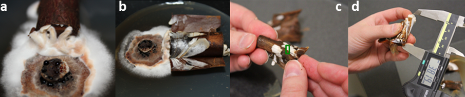
Twig and root segments colonization assay (above). From left to right, a stem/root segment is placed on an actively growing colony of Armillaria (a, b); colonization of the tissue after peeling back the periderm (white coloration is the Armillaria mycelial fan) (c); infected tissue measurement (d).

The third objective addresses the need to identify anti-Armillaria compounds in two known ARR resistant Prunus rootstock species, P. cerasifera and P. munsoniana, to contribute toward the breeding of ARR resistant rootstocks. Labs at Michigan State University are evaluating plant material suspected of being resistant using a twig and root colonization assay.
To seek new ARR resistance sources, the Gasic lab (Clemson University) adopted an in vitro screening protocol (Baumgartner et al. 2018) to evaluate 81 Prunus accessions, representing 28 species, from the USDA-ARS National Clonal Germplasm Repository in Davis, CA.
Microscopy is used to confirm the fungal presence and penetration from accessions that appear to be tolerant. Pictured to the right is a 40x magnification of the root (red) and fungus (green). A susceptible accession (left) displays fungal root penetration while the resistant genotype (right) shows no fungal penetration.
References:
Warnstrom, E. L., Outwater, C. A., Jacobs, J. L., & Hammerschmidt, R. (2011). Development of an in vitro bioassay to screen Prunus spp. for resistance to Armillaria ostoyae. Phytopathology, 1(6).
Kaniszewski, L. J., Hammerschmidt, R. (2015). Novel phytoanticipins from Prunus maackii: Possible factors in disease resistance. Michigan State University.
Baumgartner, K., Fujiyoshi, P., Ledbetter, C., Duncan, R., & Kluepfel, D. A. (2018). Screening Almond Rootstocks for Sources of Resistance to Armillaria Root Disease. HortScience, 53(1), 4-8.
In Vitro Screening
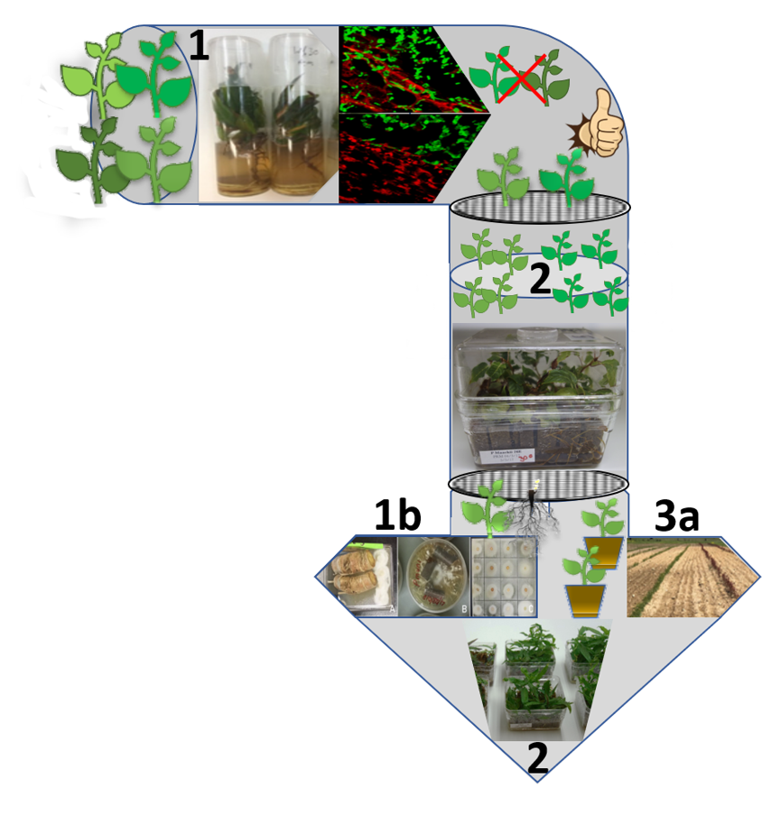
In vitro screening protocol to assess the susceptibility/tolerance of Prunus genotypes to Armillaria was developed. Prunus germplasm in vitro screening developed by Baumgartner et al. (2018) and root microscopy confirmation of absence of fungal penetration are used in Step 1. Resistant germplasm from Step 1 is multiplied and grown in co-culture with Armillaria in Oasis® IVE phenolic resin (Step 2). Germplasm with confirmed resistance after in vitro screen 1 and 2 is multiplied and analyzed for anatomical and biochemical (Objective 1b), and transcriptomic and metabolomic (Objective 2) responses to infection. Stocks of healthy plants are maintained in vitro, in pots in the greenhouse and field for assessing optimal horticultural characteristics (Objective 3a).
Using the new resistance screening pipeline we have tested 81 Prunus spp accessions from Prunus National Clonal Germplasm Repository (NCGR) in Davis, CA and 4 from Clemson Prunus germplasm for their response to ARR infection in vitro.
Resistant response, observed in 4 accessions in vitro was confirmed by root microscopy.
Antifungal activity of the compounds from the root periderm of 28 different Prunus genotypes was investigated using three-step, in vitro antifungal activity screening bioassay.
Antifungal compounds phytoanticipins were detected, specifically flavonoids, including uncommon flavonoids, alnusin and alnustinol and their precursors.
We demonstrated host’s ability to limit ARR infection by several collective nonspecific host responses acting together, such as the formation of new callus tissue on the root surface, a colored reaction zone, necrophylactic periderm, new cells, and new vascular cambium to compartmentalize the pathogen.

Mode of tolerance/resistance in Prunus. a, c – Necrophylactic periderm (np) formation; b – Intact bark walling off; d – A. mellea infection spreading around the bark of S-37 Stribling and thin layer of newly formed cells (nc) unable to compartmentalize the fungal spread and infection; e – Healthy wounded root of P. mahaleb #1 with internal callus (ic) formation around the cambial region; f – D. tabescens infection in P. mahaleb #1 root. Bars = 200 µm (a, b, d, e, and f) and 50 µm (c) (Devkota et al., 2020)
Devkota, P., Iezzoni, A., Gasic, K., Reighard, G., and R. Hammerschmidt (2020) Evaluation of the susceptibility of Prunus rootstock genotypes to Armillaria and Desarmillaria species. European Journal of Plant Pathology. https://doi.org/10.1007/s10658-020-02065-y