Objective 1– Identify, Characterize, and Utilize Sources of Resistance to Armillaria /Desarmillaria
Summary of Objective 1
Prunus germplasm exhibits various modes of resistance to Armillaria root rot (ARR). Prunus breeders collaborating with pathologists will evaluate germplasm from four sources (nurseries, USDA-ARS NCGR, wild collection, and hybrids from directed crosses) for resistance to Armillaria solidipes, A. mellea or Desarmillaria tabescens by implementing a resistance screening pipeline (Fig below). All resistant germplasm will be further characterized for its anatomical and biochemical response to infection and used in breeding and field evaluation
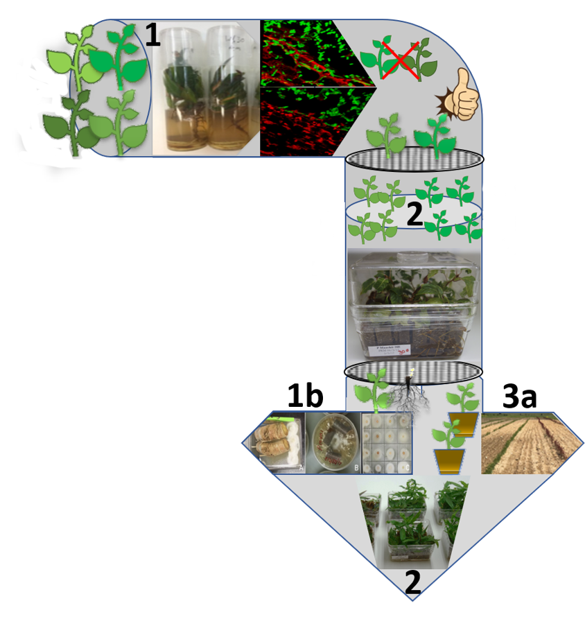
Figure 1 Resistance screening pipeline. Prunus germplasm in vitro screening (Baumgartner et al. 2018) and root microscopy confirmation of absence of fungal penetration (Step 1). Resistant germplasm multiplication and co-culture with Armillaria in Oasis® IVE phenolic resin (Step 2). Germplasm with confirmed resistance after in vitro screen 1 and 2 is multiplied and subjected to anatomical and biochemical (Obj. 1b), and transcriptomic and metabolomic (2) responses to infection. Stocks of healthy plants are maintained in vitro, in pots in the greenhouse and field for assessing optimal horticultural characteristics (Obj. 3a). (K. Gasic)
Obj. 1a. Identify sources of resistance in the germplasm
Team Lead: K. Gasic
Team Members: A. Iezzoni, T. Gradziel, M. Allakarjuana, P. Devkota and A. Calle
Resistance screening pipeline (Figure 1) was developed to quickly assess tolerance to Armillaria in vitro. Prunus germplasm is initially screened using in vitro protocol developed by Baumgartner et al. (2018) and root microscopy is used to confirm tolerance via absence of fungal penetration (Step 1; Figure 2). Resistant germplasm from Step 1 is multiplied and grown in co-culture with Armillaria in Oasis® IVE phenolic resin (Step 2). Germplasm with confirmed resistance after in vitro screen 1 and 2 is multiplied and analyzed for anatomical and biochemical (Objective 1b), and transcriptomic and metabolomic (Objective 2) responses to infection. Stocks of healthy plants are maintained in vitro, in pots in the greenhouse and field for assessing optimal horticultural characteristics (Objective 3a).
Using the new resistance screening pipeline we have tested 81 Prunus spp accessions from Prunus National Clonal Germplasm Repository (NCGR) in Davis, CA and 4 from Clemson Prunus germplasm for their response to ARR infection in vitro.
Resistant response, observed in 4 accessions in vitro was confirmed by root microscopy.

Figure 2. Plum (Prunus cerasifera accession DPRU.2101) roots show Armillaria mycelium (green) on the root (red) surface, but no evidence of penetration. Images are taken by confocal microscopy at 100 and 400X. (K. Gasic)
Obj. 1b. Characterize the anatomical and biochemical responses of root tissue to infection
Team Lead: R. Hammerschmidt
Team Members: K. Baumgartner, J. Adelberg, P. Devkota
Mechanisms of resistance in the germplasm are characterized (Figure 3). This knowledge will enable researchers to recognize phenotypic and genomic features, which could serve as breeding targets, as well as which germplasm can be used to develop rootstocks that exhibit more than one resistance mechanism.
Antifungal activity of the compounds from the root periderm of 28 different Prunus genotypes was investigated using three-step, in vitro antifungal activity screening bioassay.
Antifungal compounds phytoanticipins were detected, specifically flavonoids, including uncommon flavonoids, alnusin and alnustinol and their precursors.
We demonstrated host’s ability to limit ARR infection by several collective nonspecific host responses acting together, such as the formation of new callus tissue on the root surface, a colored reaction zone, necrophylactic periderm, new cells, and new vascular cambium to compartmentalize the pathogen (Figure 3).

Figure 3. Mode of tolerance/resistance in Prunus. a, c – Necrophylactic periderm (np) formation; b – Intact bark walling off; d – A. mellea infection spreading around the bark of S-37 Stribling and thin layer of newly formed cells (nc) unable to compartmentalize the fungal spread and infection; e – Healthy wounded root of P. mahaleb #1 with internal callus (ic) formation around the cambial region; f – D. tabescens infection in P. mahaleb #1 root. Bars = 200 µm (a, b, d, e, and f) and 50 µm (c) (Devkota et al., 2020)
Devkota, P., Iezzoni, A., Gasic, K., Reighard, G., and R. Hammerschmidt (2020) Evaluation of the susceptibility of Prunus rootstock genotypes to Armillaria and Desarmillaria species. European Journal of Plant Pathology. https://doi.org/10.1007/s10658-020-02065-y